All good things come to an end so the saying goes.  With that I bid you adieu. This is my last enry.

As long as I live, I will have complications from having surgeries on my brainstem.  Sure, I’ve given up a lot, but I have gained a lot, too.  For instance, I am alive and have witnessed the long term commitment of my husband; watched my grandchildren develop and try to find their place in this world; see my sons excel at being great husbands, fathers, providers, and men.  For all this I am grateful.

Beth Grigsby

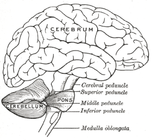
Everyone has a brainstem.  It has nothing to do with intelligence but a lot to do switch everything else.   The brainstem:  In vertebrate anatomy the brainstem (or brain stem) is posterior part of the brain, adjoining and structurally continuous with the spinal cord.


The brain stem provides the main motor and sensory innervation to the face and neck via the cranial nerves. Though small, this is an extremely important part of the brain as the nerve connections of the motor and sensory systems from the main part of the brain to the rest of the body pass through the brain stem. This includes the corticospinal tract (motore), the posterior column-medial lemniscus pathway (fine touch, vibration sensation and proprioception) and the spinothalamic tract (pain, temperature, itch and crude touch)

The brain stem also plays an important role in the regulation of cardiac and respiratory function. It also regulates the central nervous system, and is pivotal in maintaining consciousness and regulating the sleep cycle. The brain stem has many basic functions including heart rate, breathing, sleeping and eating. It is usually described as including the medulla oblongata (myelencephalon), pons (part of metencephalon), and midbrain (mesencephalon).

Source: Wikipedia

Once a week we go to physical therapy (PT), it follows on the heels of other PT I’ve had.  We venture north to a place in Minneapolis called Courage Center.  It Is a place to go for people like me.   Their mission is:  ” to empower people with disabilities realize their  full potential in every aspect of life.  We are guided by a vision that, one day, all people will live, work, learn and play in a community based on abilities not disabilities.”

I  can’t  recommend the Courage Center enough. They help all kinds of people.   For me, it’s a humbling experience.  To see people there who breathe through a tube as well as talk, and more is heart-wrentching.  Courage Center is up to the challenge and performs admirably.

The therapist and I go through exercises to strengthen the muscles in my back.  We are  also working on my posture.  A lot of it comes from my pons, where I had my second surgery.  My core muscles are in good condition so we work primarily on the back.

I feel it is important to continue exercising at home, after PT.  Not just at PT.  So, I have a regular program that mimics PT and takes me about 20 minutes everyday.  Even if you are in good health I urge you to get some form of exercise.

Source: Courage Center literature

Since I had surgery on The Pons area of my brainstem it is a very familiar place.  I wanted Wikipedia’s take, too,  Here it is:

The pons is a structure located on the brain stem, named after the Latin word for the 16th-century Italian anatomist and surgeon Costanzo Varolio (pons Varolii).    It is cranial to the medulla oblongata, caudal to the midbrain, and ventral to the cerebellum. In humans and other bipeds this means it is above the medulla, below the midbrain, and anterior to the cerebellum. This white matter includes tracts that conduct signals from the cerebrum down to the cerebellum and medulla, and tracts that carry the sensory signals up into the thalamus.

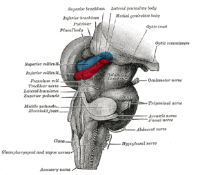
The pons measures about 2.5 cm in length. Most of it appears as a broad anterior bulge rostral to the medulla. Posteriorly, it consists mainly of two pairs of thick stalks called cerebellar peduncles[disambiguation needed]. They connect the cerebellum to the pons and midbrain.

The pons contains nuclei that relay signals from the forebrain to the cerebellum, along with nuclei that deal primarily with sleep, respiration, swallowing, bladder control, hearing, equilibrium, taste, eye movement, facial expressions, facial sensation, and posture,

Within the pons is the pneumotaxic center, a nucleus in the pons that regulates the change from inspiration to expiration.

The pons is implicated in sleep paralysis, and also plays a role in generating dreams.


     We were walking the neighborhood.  He on foot, I on my electric wheelchair, when Jeff looked and saw a woman.  He called to her to get her attention.  He had noticed the recumbent bicycle in her garage.  We wanted to try one to see if I could do it.  After a little discussion and trying, low and behold, I could.

It turns out that she wanted to sell it, she would also let us use it for a week to try it out.  We took her up on her offer and bought it from her after a few days.  So far, it has done well.  Jeff straps me in, and off we go.

I haven’t had cardiovascular exercise for years.  Bicycle riding is great.  When I was normal, I loved riding.   Since my last surgery, I had to give that up since I have no balance.  That’s why a tandem, recumbent bicycle works so well for me. No balance is required.  I can’t tell you how wonderful the wind feels and to have a sense of freedom is almost overwhelming.

     Last week, there was a cancellation in the appointments of the neuro opthmogist.  We were offered the slot and took it.  I am concerned about my right eyelid closing and hoped the specialist could suggest a course of treatment to aid me, other than surgery.

     We waited our turn in a long, L-shaped waiting room, typical of most, with the receptionist tucked neatly behind her desk.  Before long, my name was called, and we went in.

     No matter what kind of doctors we see, I am always nervous. I think it has to do with my mortality, which I don’t like to think about.

     To make a long story short, here are the results of our visit:

     There is, as we suspected, nothing we can do.  As far as my right eye is concerned, nature is doing its thing by closing. It is helping me with my double vision.  Right now I only use one eye to see.  The right lens of my glasses is etched so I cannot see using that eye. It makes seeing impossible, thus, one has to use the other eye.

     Then, we talked about that eye being firmly in the far corner of my right eye.  Apparently, I have damage to my optical nerve.  Any surgery, which I don’t want, would result in a short-term fix.   Eventually, the right eye would drift back to the corner, the long-term position.

     We left, and I was downhearted. Oh well, so it goes.

     Thank you for giving me this opportunity to share my AVM experiences with you.  It was very helpful.  Getting over this will take years, if ever, and every year is better.  The challenges remain, and continue to be a struggle.  I met many good people along the way and saw extra goodness in others.  I am very grateful.

     Synapses are the key functional elements of the brain.  The essential function of the brain is cell-to-cell communication, and synapses are the points at which communication occurs. The human brain has been estimated to contain approximately 100 trillion synapses; even the brain of a fruit fly contains several million. The functions of these synapses are very diverse: some are excitatory (excite the target cell); others are inhibitory; others work by activating second messenger systems that change the internal chemistry of their target cells in complex ways.  A large fraction of synapses are dynamically modifiable; that is, they are capable of changing strength in a way that is controlled by the patterns of signals that pass through them. It is widely believed that activity-dependent modification of synapses is the brain’s primary mechanism for learning and memory.

      In short, synapses occur constantly and jump from cell to cell in our brains.  You might find the following interesting:    

      The human brain is the center of the human nervous system. It has the same general structure as the brains of other mammals, but is larger than expected on the basis of body size among other primates.  Estimates for the number of neurons (nerve cells) in the human brain range from 80 to 120 billion.  Most of the expansion comes from the cerebral cortex, especially the frontal lobes, which are associated with executive functions such as self-control, planning, reasoning, and abstract thought. The portion of the cerebral cortex devoted to vision is also greatly enlarged in human beings, and several cortical areas play specific roles in language, a skill that is unique to humans.

      Despite being protected by the thick bones of the skull, suspended in cerebrospinal fluid, and isolated from the bloodstream by the blood–brain barrier, the human brain is susceptible to many types of damage and disease. The most common forms of physical damage are closed head injuries such as a blow to the head, a stroke, or poisoning by a variety of chemicals that can act as neurotoxins. Infection of the brain, though serious, is rare due to the biological barriers which protect it. The human brain is also susceptible to degenerative disorders, such as Parkinson’s disease, multiple sclerosis, and Alzheimer’s disease. A number of psychiatric conditions, such as schizophrenia and depression, are thought to be associated with brain dysfunctions, although the nature of such brain anomalies is not well understood.

      Researchers who study the functions of the cortex divide it into three functional categories of regions, or areas. One consists of the primary sensory areas, which receive signals from the sensory nerves and tracts by way of relay nuclei in the thalamus. Primary sensory areas include the visual area of the occipital lobe, the auditory area in parts of the temporal lobe and insular cortex, and the somatosensory area in the parietal lobe. A second category is the primary motor area, which sends axons down to motor neurons in the brainstem and spinal cord. This area occupies the rear portion of the frontal lobe, directly in front of the somatosensory area. The third category consists of the remaining parts of the cortex, which are called the association areas. These areas receive input from the sensory areas and lower parts of the brain and are involved in the complex process that we call perception, thought, and decision making.

     There is a whole lot online and other places about this very complex organ. 

Source:  Wikipedia

     Two surgeries on my brain stem have made me interested in the brain and its workings.  What follows is not a tutorial but more of a refresher of what you had in your Human Biology course in high school.

      The brain is a complex organ.  It sits in our head and is composed of tissue.  The brain is responsible for our sight, hearing, smell, touch, taste, balance, all the senses.  It is the center of our nervous system.  We, as human beings, are mammals and vertebrates.  We have brains central to our development and well-being.

      The different areas of our brain are:   The medulla, along with the spinal cord, contains many small nuclei involved in a wide variety of sensory and motor functions.

     The pons lies in the brainstem directly above the medulla. Among other things, it contains nuclei that control sleep, respiration, swallowing, bladder function, equilibrium, eye movement, facial expressions, and posture.

      The hypothalamus is a small region at the base of the forebrain, whose complexity and importance belies its size. It is composed of numerous small nuclei, each with distinct connections and neurochemistry. The hypothalamus regulates sleep and wake cycles, eating and drinking, hormone release, and many other critical biological functions.

      The thalamus is another collection of nuclei with diverse functions. Some are involved in relaying information to and from the cerebral hemispheres. Others are involved in motivation. The subthalamic area (zona incerta) seems to contain action-generating systems for several types of “consummatory” behaviors, including eating, drinking, defecation, and copulation.

      The cerebellum modulates the outputs of other brain systems to make them precise. Removal of the cerebellum does not prevent an animal from doing anything in particular, but it makes actions hesitant and clumsy. This precision is not built-in, but learned by trial and error. Learning how to ride a bicycle is an example of a type of neural plasticity that may take place largely within the cerebellum.

      The optic tectum allows actions to be directed toward points in space, most commonly in response to visual input. In mammals it is usually referred to as the superior colliculus, and its best-studied function is to direct eye movements. It also directs reaching movements and other object-directed actions. It receives strong visual inputs, but also inputs from other senses that are useful in directing actions, such as auditory input in owls and input from the thermosensitive pit organs in snakes. In some fishes, such as lampreys, this region is the largest part of the brain. The superior colliculus is part of the midbrain.

      The pallium is a layer of gray matter that lies on the surface of the forebrain. In reptiles and mammals, it is called the cerebral cortex. Multiple functions involve the pallium, including olfaction and spatial memory. In mammals, where it becomes so large as to dominate the brain, it takes over functions from many other brain areas. In many mammals, the cerebral cortex consists of folded bulges called gyri that create deep furrows or fissures called sulci. The folds increase the surface area of the cortex and therefore increase the amount of gray matter and the amount of information that can be processed.

      The hippocampus, strictly speaking, is found only in mammals. However, the area it derives from, the medial pallium, has counterparts in all vertebrates. There is evidence that this part of the brain is involved in spatial memory and navigation in fishes, birds, reptiles, and mammals.

      The basal ganglia are a group of interconnected structures in the forebrain. The primary function of the basal ganglia appears to be action selection: they send inhibitory signals to all parts of the brain that can generate motor behaviors, and in the right circumstances can release the inhibition, so that the action-generating systems are able to execute their actions. Reward and punishment exert their most important neural effects by altering connections within the basal ganglia.

      The olfactory bulb is a special structure that processes olfactory sensory signals and sends its output to the olfactory part of the pallium. It is a major brain component in many vertebrates, but is greatly reduced in primates.

Source: Wikipedia

      I hope you enjoyed this because there is more coming,
Our thoughts and prayers are focused on the great losses in Colorado.

We went to two museums in the last week.  One is called Traverse de Sioux the other The Mill City Museum.  Both run by the Minnesota Historical Society.

At the Traverse de Sioux museum, we learned more about the early settlers and the Sioux uprising here in Minnesota.  It certainly was a tumultuous time. Did you know people came here by steam-driven riverboats by the hundreds just to settle here.

At the Mill City Museum, we learned a little about flour production.  Mill City is a nickname given to Minneapolis because there were so many flour mills along the great Mississippi River during the 1800’s.

Both were really good museums,  especially if you like history.

What I am really saying is life doesn’t stop when there is a condition.  Yes, it changes, but it’s there for the taking.  Sure, I wish I could do these things without my wheelchair  but I cannot.  I do try to enjoy them, though.